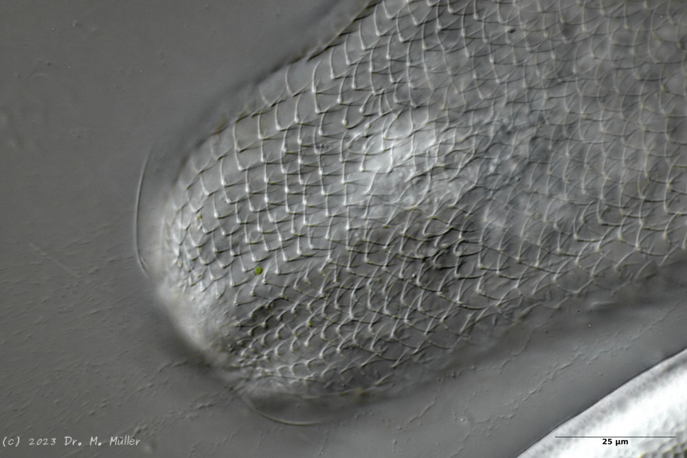
The genus Aspidiophorus is very easily recognized microscopically by the apparent “double contour” of the body. This double contour is produced by the typical style scales of this genus, which consist of a base plate that sits directly on the cuticle. From this base plate rises a thin peduncle, which carries at its end a - usually larger - un-spined terminal plate:
typical scales of an Aspidiophorus, here A. squamulosus.
This results in the typical double outer contour of the animals:
Diagnosis
Body bottle shaped
Furka normal
Peduncle scales
Subsections of Aspidiophorus
Aspidiophorus aster
Taxonomy
Order: CHAETONOTIDA Remane, 1925
Suborder: PAUCITUBULATINA d'Hondt, 1971
Family: CHAETONOTIDAE Gosse, 1864
Subfamily: CHAETONOTINAE Kisielewski, 1991
Genus: Aspidiophorus Voigt, 1903
Species: aster
Length ( bottle ):
Width:
Width of the head ( five-lobed ):
µm
Length of the furca:
Adhessive tubes:
Pharyx ( cylindrical ):
Diameter of the mouth ( around ): unknown
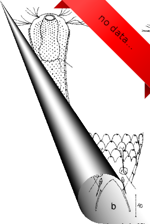
Unfortunately, I have no data available for this species!
If you have access to the following literature:
Martin,L.V. 1981. Gastrotrichs found in Surrey. Microscopy 34: 286-300.
I would be very happy if you would send me a copy by Mail!
Unfortunately, I have no data available for this species!
If you have access to the following literature:
Grosso,L.E. 1973. Notas sobre Gastrotrichos Argentinos II. Neotropica 19(59): 87-89.
Grosso,L.E. 1973. Notas sobre Gastrotrichos Argentinos I. Chaetonotus (Zonochaeta) guruguetoi sp. nov. y Aspidiophorus brahmsi sp. nov. Physis 32B: 133-137.
I would be very happy if you would send me a copy by Mail!
Width of the head ( three lobed ): 17 µm - 18.5 µm
µm
Length of the furca: 20 µm
Adhessive tubes: 50% of furca
Pharyx: 32 µm - 36 µm
Diameter of the mouth ( around ): 5.5 - 8.5 µm
Dorsal scales: rhombic petiole scales (3µm long), 41-42 per row, last row in middle with spines (9-14µm), laterally at base of toes a pair of long spines (16-18µm), sometimes with another pair of spines or elongated scales
Ventral scales: Interciliary field with a pair of terminal spines; very many minute keels
Unfortunately, I have no data available for this species!
If you have access to the following literature:
Grosso,L.E. and Drahg,F. 1983. Gastrotricos dulceacuicolas de la provincia de Tucman. I. Chaetonotus soberanus sp. nov. y Aspidiophorus lilloensis sp. nov. Neotropica 29: 189-193.
I would be very happy if you would send me a copy by Mail!
Pharyx ( cylindrical, with small terminal swellings ): 30 µm - 35 µm
Diameter of the mouth ( around ): 3 µm
Dorsal scales: 9 rows of 16-17 peduncle scales each; end plates (6-7 x 2-3 µm) rounded anteriorly, incised posteriorly, no keels; last 2-3 rows terminate in 20 µm spines; lateral on toes a pair of spines (25 µm).
Ventral scales: Ventral intercilliary field with 25 rows of tiny petiole scales
Oecology: Mud dweller
Similar species: A. longichaetus
: Head rather trilobate, no median spines
Dorsal scales: 41-44 rows of 48-50 small peduncle scales each; end plates elliptical, acuminate with keel, overlapping, 3 x 1.5 µm; two 5x2 µm peduncle scales on furca inner side.
Ventral scales: Ventral innercilliary field with two terminal plates (7x4 µm) and 10 rows of narrow, distally pointed keel scales (4x0.75µm)
Dorsal scales: 25 rows of 40-45 rhombic petiolar scales each with lateral and median keels (4x6 µm); petioles 6-7 µm; terminal plates of last terminal rows often enlarged (12-17 µm); posterior end free; between toes 4 short spines on oval scales.
Ventral scales: Ventral innerciliary field covered with petiole scales
Oecology: Sludge dweller
Similar species: distinctive, variable type
Particularities: 3 teeth, eggs smooth (125x74 µm)
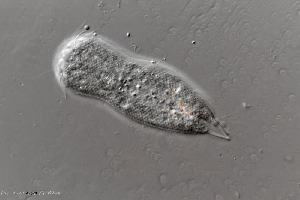
Aspidiophorus paradoxus is the largest Aspidiophorus species with about 300µm length.
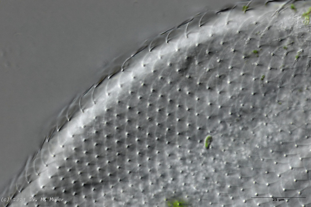
Dorsal scales
The entire animal is covered with relatively large rhombic peduncle scales.
Cross section
In cross-section, the structure of the petiole scales is clearly visible: the scales sit with a small base plate on the cuticle of the animals, from which rises a thin, hollow pedicel. At the end of the peduncle sits a rhombic terminal plate, with a central keel. At the posterior end of the animal, the terminal plates of the last row of scales are enlarged.
The pharynx of the animal is terminally swollen, and the head is weakly five-lobed with two separate pairs of palpal tufts.
Central view
Ventrally, the strong hypostomion behind the mouth opening is striking. The two ciliated bands split at the head, but the inner branches do not unite in the population I examined. The base of the toes does not bear scales, the adhesive tubes
measure about 50% to 70% of the toe length and taper to a point.
Let’s take a closer look at the scales:
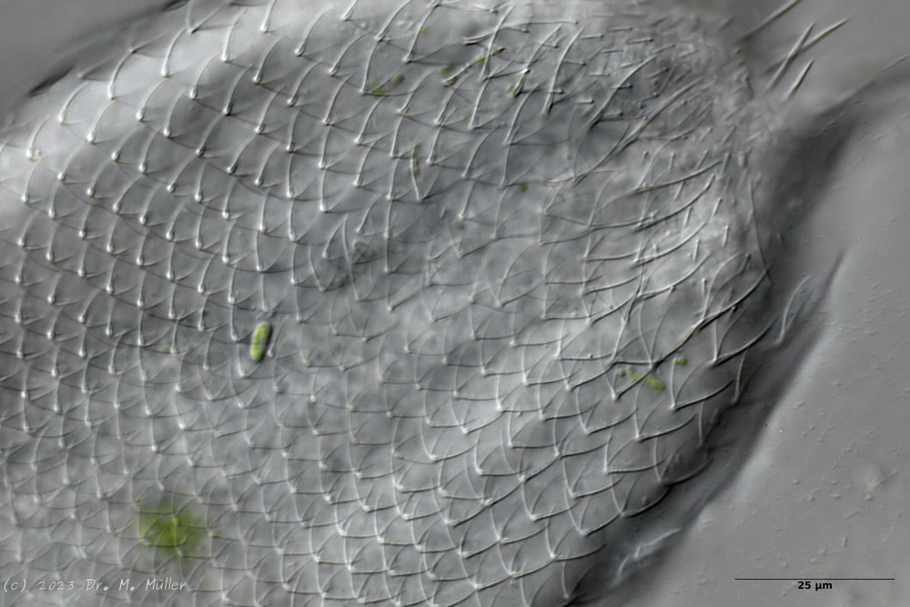
Backscales
In the scales on the back, the rhombic shape of the end plates is most clearly recognizable. Less conspicuous - but typical for the species - is the central keel of the scales.
Cross section shed
In cross-section, the complex geometry of the stem scales becomes clear - base plate, stem and end plate form a very flexible and stable armor. The additional cavity under the outer scales acts like a “crumple zone” and further increases the protective effect.
Cross section scale stems
The stems of the scales consist of hollow tubes that ensure maximum stability with minimum material input - a fascinating example of evolutionary optimization.
The abdomen of the animals is not completely covered with petiolar scales. Rather, they end in the anal region and are joined by simple small, rounded keel scales that are not an obstruction to feces.
Scaling of the abdomen.
At the base of the furca some (according to literature 4) spines protrude into the toe.
The head is almost completely covered with slightly smaller peduncle scales:
Headshed
Cephalion and pleurae are quite small and inconspicuous.
According to literature A. paradoxus has three teeth in the pharynx:
Mouth armament
In the animals I examined, there was only one curved stylet brace, the tip of which protrudes into the lumen of the pharynx and is probably used to open algal cells that are conveyed past it. It is possible that the literature reference to “three teeth” is merely based on a microscopic artifact, as the entire clasp may not be in the focal plane as a whole.
Dorsal scales: 45-53 rows of 29-32 petiole scales each (5-7 µm); no keel, not rhombic, similar shape to that of
P. rhomboides
; Toe base dorsal with keels
Ventral scales: 6 keeled terminal plates; at the posterior end 10-12 rows of keels (4-4.5 µm), otherwise naked
Oecology: between roots; Brazil
Particularities: very small pharynx; body very broad
Dorsal scales: 18-20 rows of 42-45 small petiolar scales each; petioles 1.5 µm; terminal plates 2-3.5 µm long, proximally strengthened margin, distally acuminate, weak median keel; no spines
Ventral scales: Ventral intercilliary field naked, except for a few elongate peduncle scales; two narrow terminal plates(6.5-11.5 µm)
Oecology: Bog, mud
Similar species: delimited by naked ventral intercilliary field
Particularities: without pseudocells; naked ventral intercilliary field .
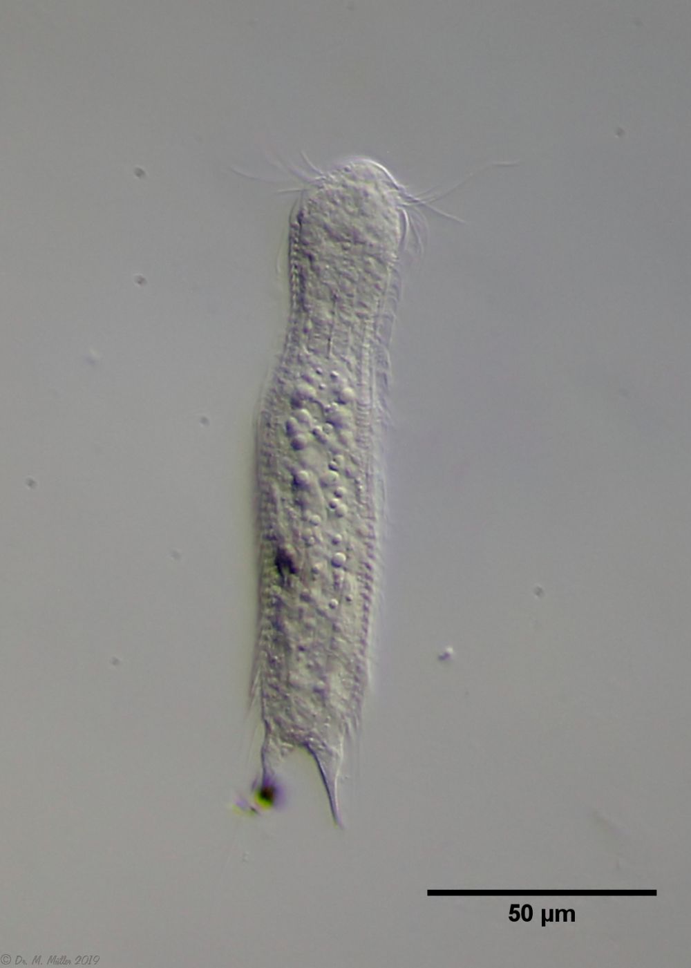
I have found this belly-harper only once so far in a bog (Sima bog). By the naked ventral intermediate field with long terminal plates the species is well to delimitate:
The round anterior margins of the end plates of the lateral peduncle scales are also well visible. The found animal is with ca. 130 µm a little smaller than the literature specification and all other measurements scale accordingly. The mouth ring is very small and strongly marked. A hypostomium does not exist.
In the median optical section the dumbbell-shaped pharynx can be seen well. The scales are distally long extended (unfortunately the shape of the end plates could not be seen). The granular structure of the two adhesive glands is also interesting.
Width of the head ( five-lobed, pointed cephalion ): ? µm
µm
Length of the furca: 16 µm
Adhessive tubes (tapered, claws): 50% of furca
Pharyx ( cylindrical with slight swellings ): 37 µm
Diameter of the mouth ( around ): 7 µm
Dorsal scales: 53 petiole scales (3 µm) per row; 3 pairs of spines at outer base of toes (20-22 µm).
Ventral scales: Ventral intercilliary field very many petiole scales; a pair of terminal spines (8-10 µm) and many shorter spines protruding into the toe cutout.
Oecology: Mud dweller
Similar species: well demarcated by claws and spines
Dorsal scales: 25-30 rows, each with 60-80 minute petiolar scales; terminal plates unkeeled (0.5-2 µm), elongate, pointed distally; posterior end with elongate keel scales
Ventral scales: Ventral intercilliary field numerous longitudinal rows of minute petiolar scales; two keeled terminal plates (6-7 µm).
Dorsal scales: 12-15 rows of 40-42 petiolar scales each with rhombic terminal plates without median keel (3-4 x 5-6 µm); scale shell ends in anal region; posterior end and toes naked; 3 pairs of short spines at base of toes (8-10 µm).
Ventral scales: 2 terminal keels (5-6 µm), 12 rows of keels without base plates
Oecology: Mud dweller
Similar species: A. paradoxus
: unkeeled basal plates; spination at the posterior end; mouth grafting
Of the (acc. (Schwank, 1990))
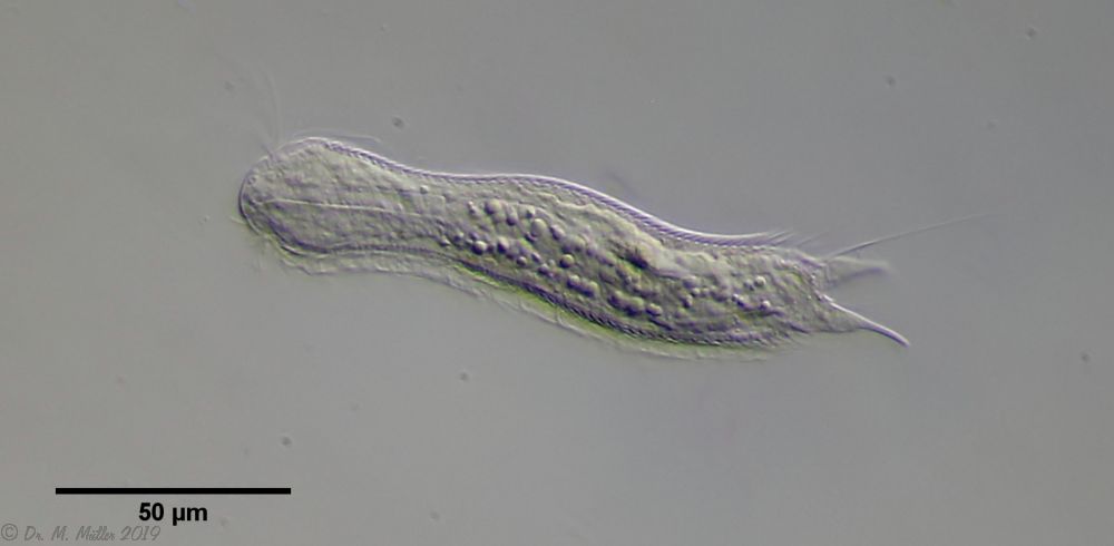
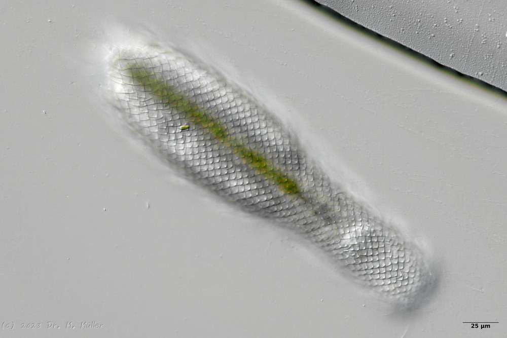
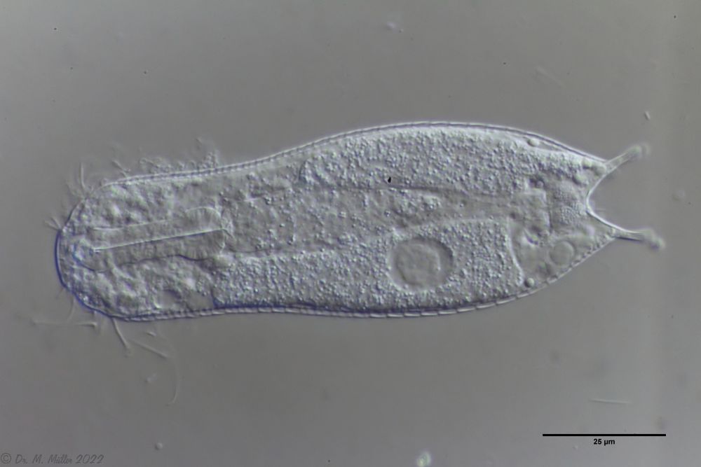
twenty limnic Aspidiophorus species only four are listed in the “official” species lists for Germany. Another species was described some time ago by Michael Plewka. The species Aspidiophorus squamulosus has been found so far only in Poland and in western France - so it is no wonder that this animal is also native to us, between the previous finding areas.
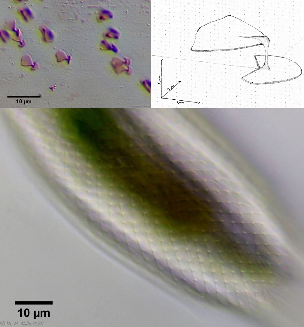
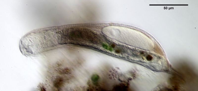
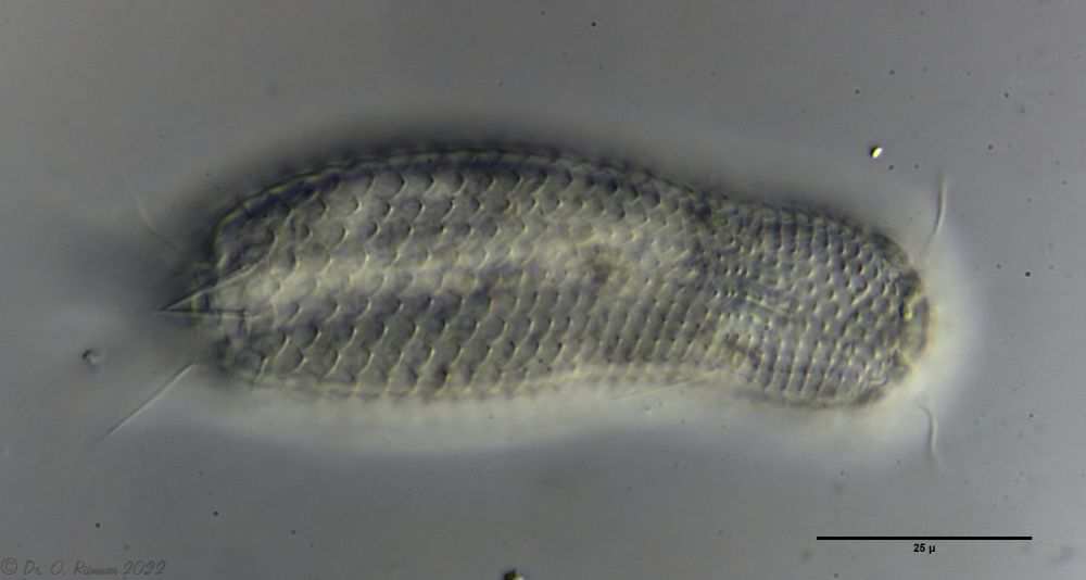
Fig. 1: Aspidiophorus squamulosus; optical section and scale image.
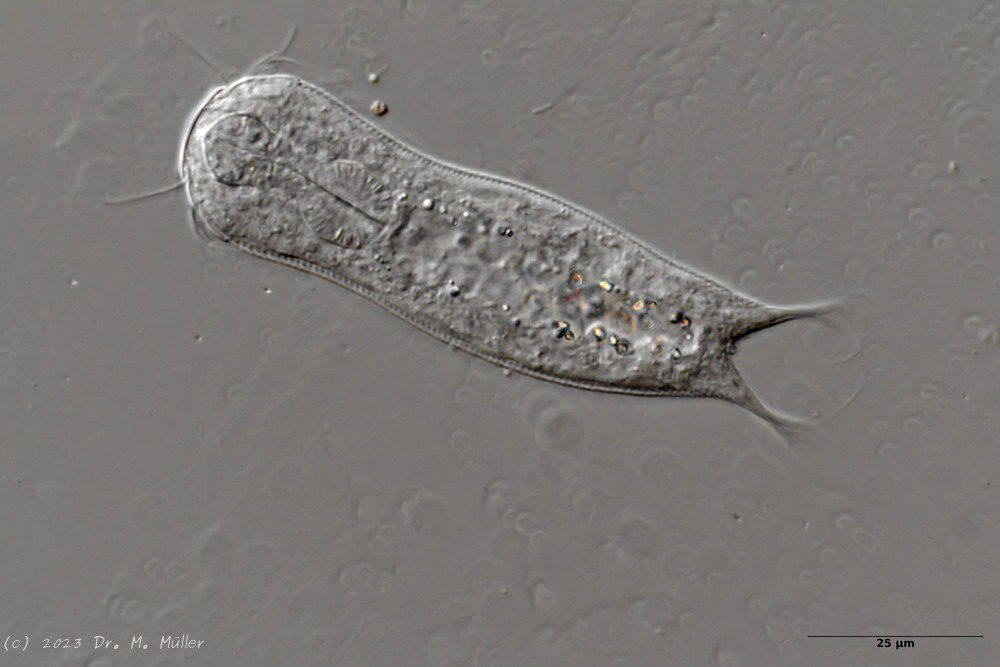
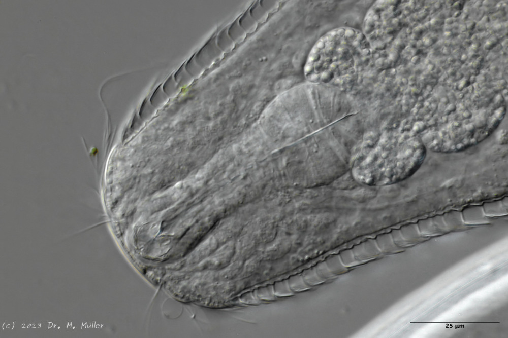
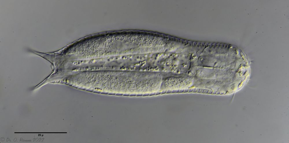
The internal structure of the animal becomes quite clear in the side view:
Fig. 2: A. squamulosus, side view.
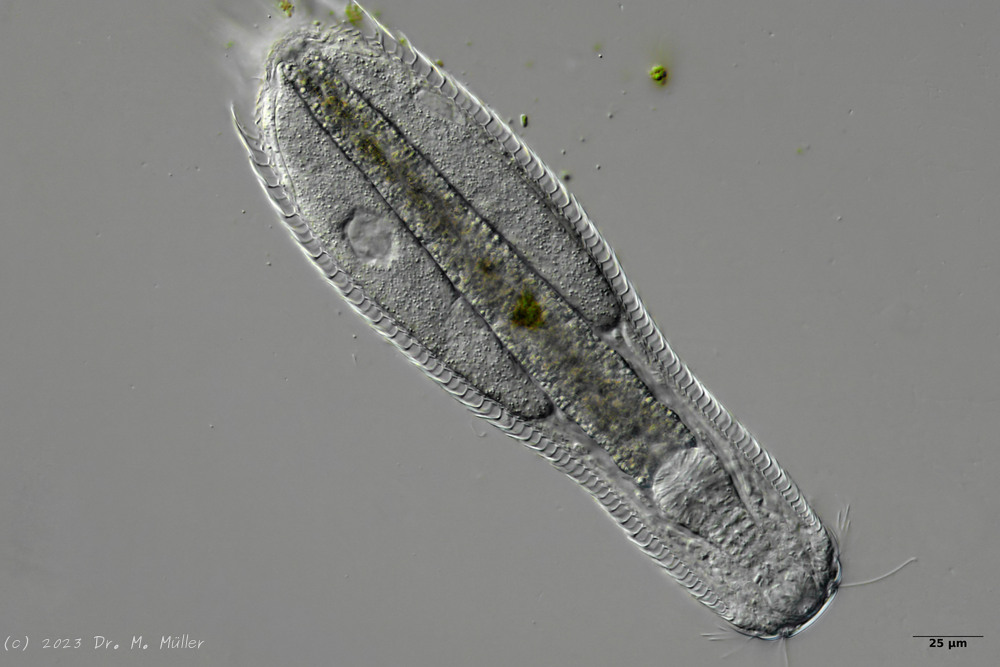
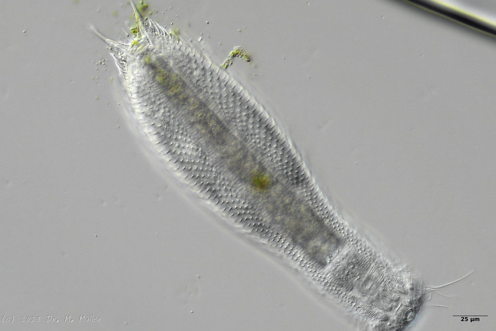
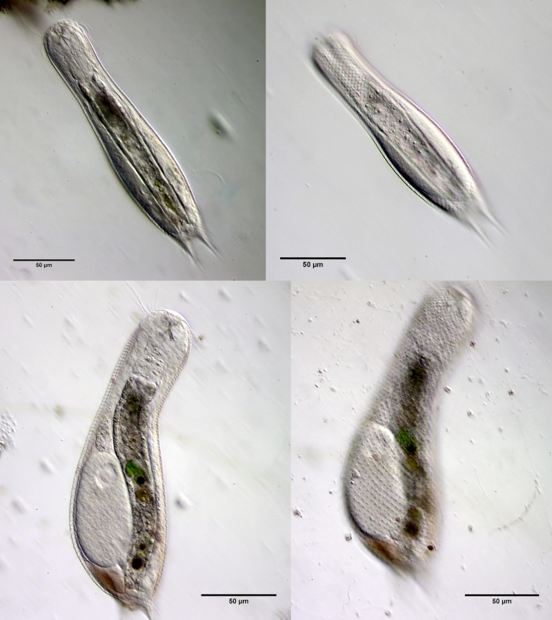
A. squamulosus is with ca. 230µm somewhat smaller than the similar species A. paradoxus and is clearly distinguished by the three spines each above the toes (pic. 3, ob. li. and mi. r.). In addition, the terminal plates of the petiolar scales do not possess a median ridge. Also the prominent, furrowed hypostomium (Fig. 3, ob. r.) is typical for this species. Some animals I found were in their “hermaphroditic phase” (post-parthenogenetic phase) and showed a very large X-organ and sperm bundle (Fig. 3, mi. l.). One animal also carried a very large egg (with a pronounced nucleolus), which it laid during the approximately 10-day observation period. Unfortunately, I was not able to observe the oviposition itself (also the egg was not found in the specimen).
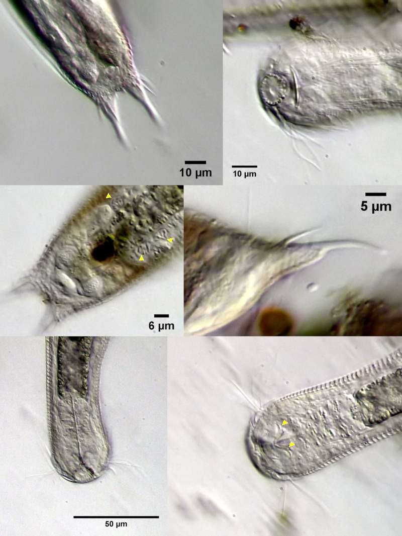
Figure 3: A. squamulosus: details;
up. l.: toe spines; up. r.: hypostomium
mi. l.: X-organ and spermathecae (marking); mi. r.: toe spines and toe scales;
low. l.: transverse section of scale carapace; low. r.: stylet (mark)_
As is often the case, there were some deviations from the species description in the literature. With up to 50 generations per year and years of isolation in a small garden pond, I find minor deviations not surprising. However, it is possible that some details were not noticed in the rare, earlier observations. For example, the base of the toes should be unscaled (Fig. 3, mi. r.). However, the specimens I observed show many very small, roundish and close-scaled scales. Only the conspicuous peduncle scales do not appear at the posterior end of the animal.
In some Aspidiophorus species movable “teeth” in the mouth tube are described. Although A. squamulosus should be toothless, two actively movable styletes could be observed, the tip of which ended in the mouth tube and reminded of tardigrades (Fig. 3, un. r.).
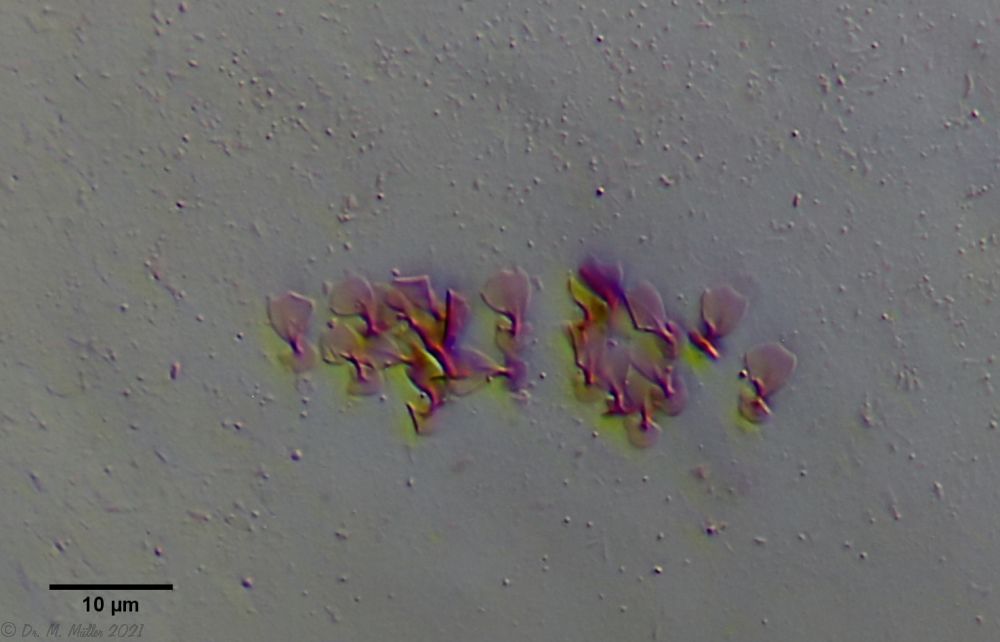
The peculiar stipe scales of the genus Aspidiophorus are not to be grasped in their three-dimensional beauty in the scale association. Therefore, I subjected a pitiable specimen of A. squamulosus to scale analysis:
Fig. 4: A. squamulosus; colored and - unfortunately - flattened single scales.
Since during a scale analysis just the very small scales are pressed rather flat, I tried a drawing reconstruction of the three-dimensional structure:
Fig. 5: A. squamulosus; bottom: composite scale image; top left: isolated and stained scales; top right: drawing reconstruction of a single peduncle scale.
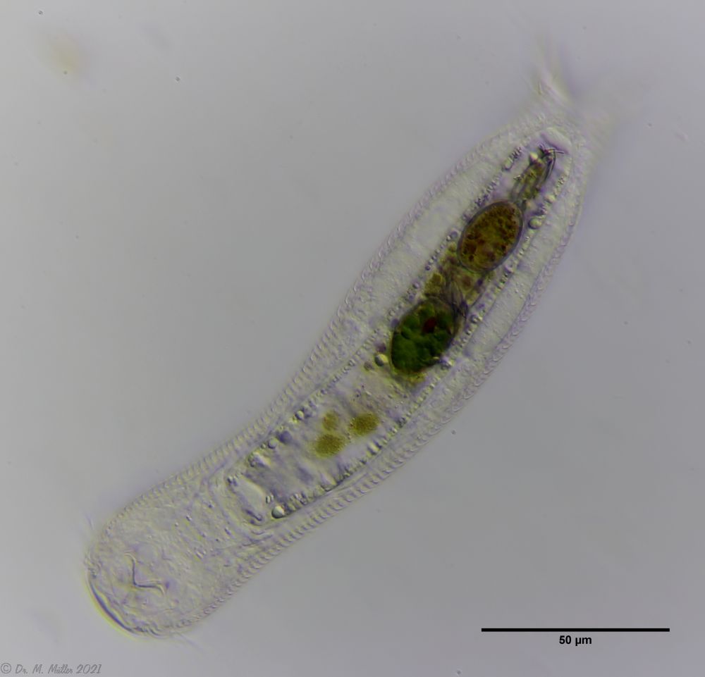
Another point that has remained open so far is the diet of A. squamulosus. The main diet consists of relatively large unicellular algae or eye flagellates. To swallow this large prey, the mouth of the animals is equipped with movable lamellae that can be folded outward for feeding (cf. Captochaetus). This allows the diameter of the mouth opening to be greatly expanded, unlike most gastrotrichs. During the swallowing movement, the stylet is pressed into the mouth tube and the prey cells are guided past it. In the process, the hard cell wall is probably perforated so that the digestive secretions in the intestine can penetrate the cells.
Fig. 6: A. squamulosus; intestine filled with ocular flagellates.
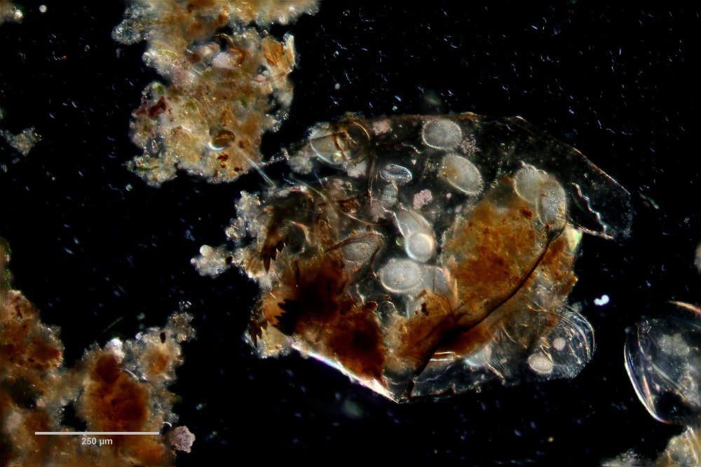
If one examines the mud of stagnant waters microscopically, one always finds gastrotrich clutches in empty shells of water fleas with sometimes dozens of eggs.
Fig. 7: A. squamulosus; focal sac of a gastrotrich clutch containing eggs of at least three different species.
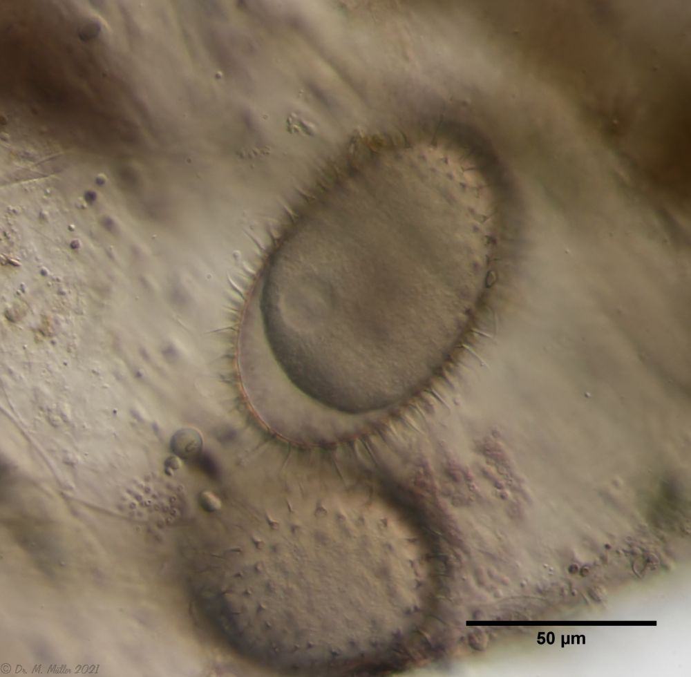
Often these clutches consist of eggs of different species. Apparently, a number of species prefer to lay their eggs in water flea shells where other gastrotrichs have already laid their eggs. In such a clutch often relatively large (95µm x 55 µm), spiny eggs are noticeable, which have not yet started embryonic development and probably represent permanent eggs.
Fig. 5: A. squamulosus; permanent egg.
One egg of the shown clutch started to develop and turned out to be an egg of the species A. sqamulosus. Therefore I can show here the eggs belonging to the species.
Dorsal scales: 15-20 rows of 38-40 very small peduncle scales each; endplates very delicate and unkeeled (2.5-3 µm); strongly bent peduncles -> scales protrude like bristles
Ventral scales: Ventral intercilliary field 9-10 rows keels; anteriorly mostly naked; no terminal plates.
Oecology: creek
Similar species: well demarcated by protruding scales
Particularities: protruding scales; body cylindrical
Dorsal scales: 15-17 rows of 33-40 petiole scales each; end plates unkeeled 2-5µm, rounded distally and proximally; base of toes naked; 2 pairs of thick spines (8-10µm and 4-5µm) on oval scales with two keels.
Ventral scales: 2 terminal keels (7.5-11µm); in gut region 8-9 rows of tiny keels, otherwise naked
Oecology: montane lake
Similar species: delimitable by spines
Particularities: 2 pairs of spines at the rear end
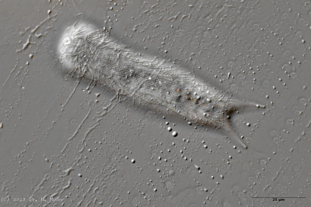
A. tetrachaetus is a very rare gastrotrich, so far confirmed only for Poland.
A. tetrachaetus: dorsal ((c) Ole Riemann)
The dorsum of the animals is covered with relatively large, unkeeled and rounded pedunculate scales, which do not continue to the base of the toes at the posterior end. A pair of large spines is conspicuous at the posterior end of the scale coat.
A. tetrachaetus: t.s. ( Ole Riemann)
In the cross section the structure of the petiole scales can be seen well.
A. tetrachaetus: ventral (copyright: Ole Riemann)
The ventral intermediate field of the animals is covered only in the intestinal region by 8-9 longitudinal rows of small keels. The pharyngeal region is naked. Two terminal keels form the posterior shoot of the ventral scaling.
A. tetrachaetus: two pairs of spines at the hind end.
Only a closer look at the dorsal toe base of the animals reveals two additional, smaller spines.
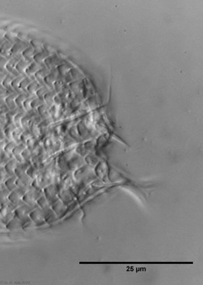
A. tetrachaetus: single tooth in oral cavity.
Contrary to the description in the literature, a single tooth can be seen protruding asymmetrically from the oral cavity in the animals at Sima Moor.
 typical scales of an Aspidiophorus, here A. squamulosus.
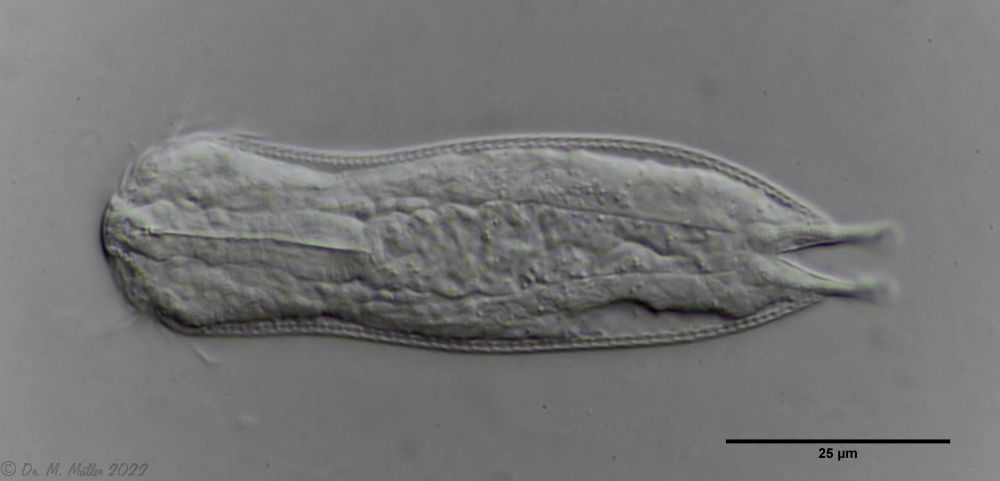


 Backscales
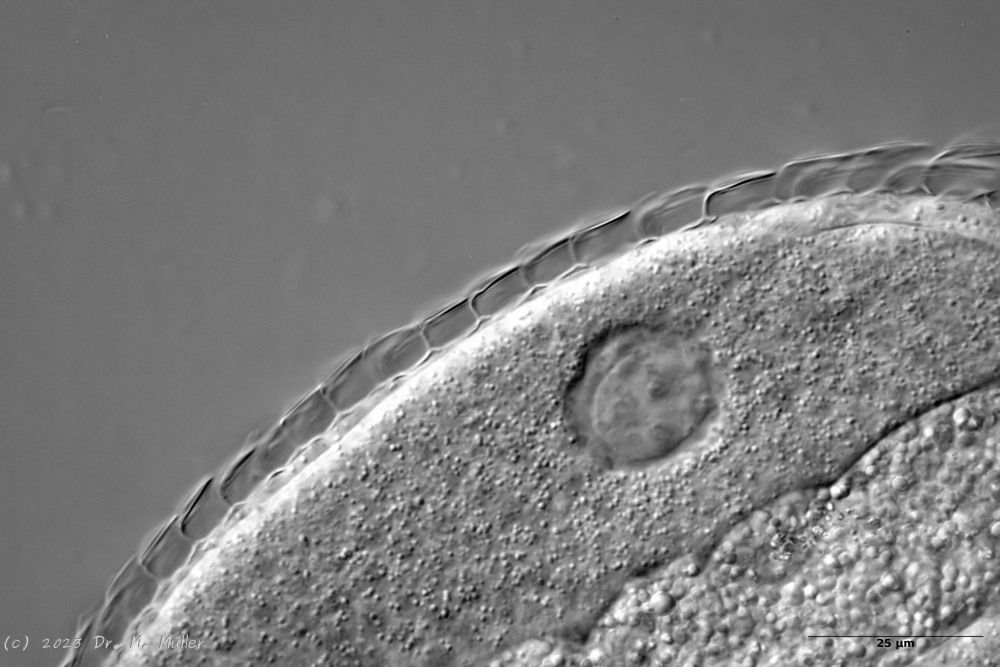
 Cross section scale stems
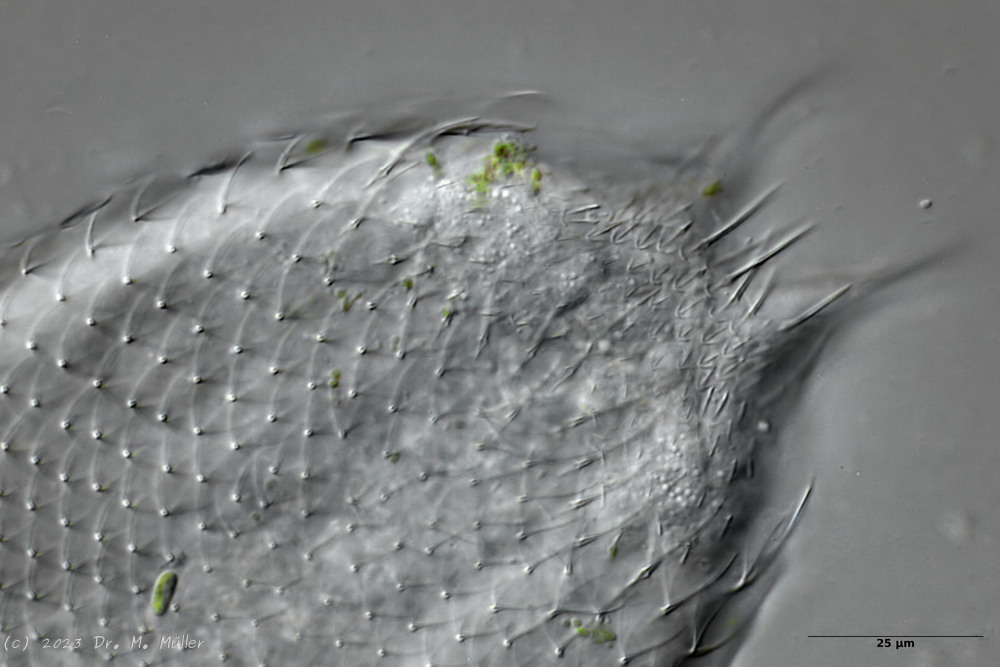


