Aspidiophorus polonicus

158 µm - 172 µm
Width:
Width of the head ( five-lobed ):
µm
Length of the furca:
16 µm - 23 µm
Adhessive tubes (thin, pointed):
50% of furca
Pharyx ( dumbbell-shaped ):
36 µm - 42.5 µm
Diameter of the mouth ( around ):
4 µm
Dorsal scales:
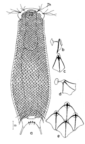
18-20 rows of 42-45 small petiolar scales each; petioles 1.5 µm; terminal plates 2-3.5 µm long, proximally strengthened margin, distally acuminate, weak median keel; no spines
Ventral scales:
Ventral intercilliary field naked, except for a few elongate peduncle scales; two narrow terminal plates(6.5-11.5 µm)
Oecology:
Bog, mud
Similar species:
delimited by naked ventral intercilliary field
Particularities:
without pseudocells; naked ventral intercilliary field .
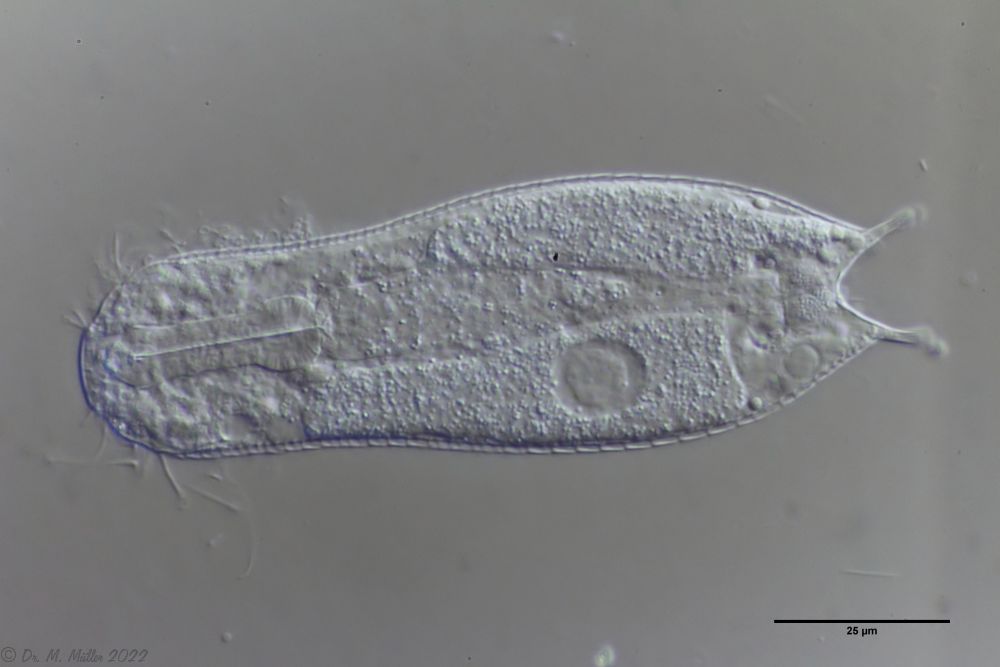
I have found this belly-harper only once so far in a bog (Sima bog). By the naked ventral intermediate field with long terminal plates the species is well to delimitate:

The round anterior margins of the end plates of the lateral peduncle scales are also well visible. The found animal is with ca. 130 µm a little smaller than the literature specification and all other measurements scale accordingly. The mouth ring is very small and strongly marked. A hypostomium does not exist.

In the median optical section the dumbbell-shaped pharynx can be seen well. The scales are distally long extended (unfortunately the shape of the end plates could not be seen). The granular structure of the two adhesive glands is also interesting.