
Scale preparation
With belly-harriers the species classification is traditionally done by the shape of the scales. Therefore, species identification is often only possible if one can accurately assess the scale shape of the animals. Unfortunately, the scales of gastrotrichs are often difficult to see. Even with DIC - although here the weak birefringence of the scales further enhances the contrast - live observation is not sufficient for species diagnosis in many species. Therefore I would like to present here a possibility for the preparation of the scales and thus for the (mostly) safe species diagnosis.
As an example I have chosen
Lepidochaetus zelinkai
. Due to the distinctive appearance of this gastrotrich, a scale analysis for species identification is actually not necessary, but some scale details are controversially discussed in the literature, so I wanted to make up my own mind.
L. zelinkai is a medium sized gastrotrich with a distinctive spiny coat:
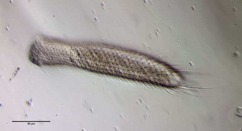
L. zelinkai
: Overview
Even with higher magnification not all details of the scaling become clear:
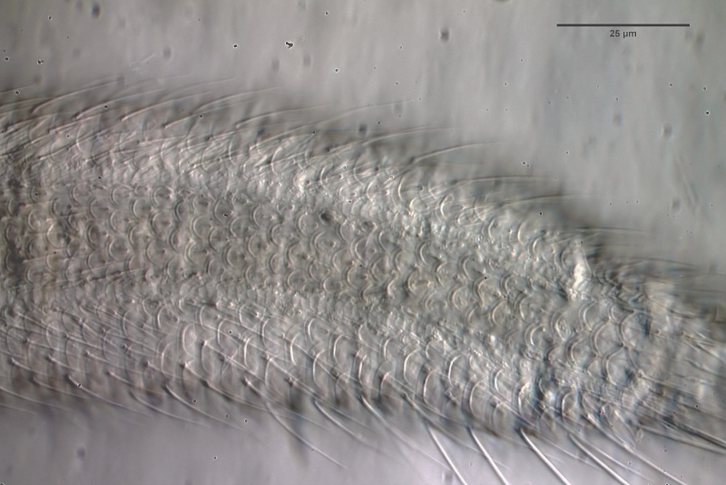
L. zelinkai
: Scales of the ventral interfield
To prepare the scales free, the animal must be macerated. To do this, fix the animal - preferably with a simple Vaseline copressorium - with the coverslip and draw a drop of glacial acetic acid under the coverslip. I stained the glacial acetic acid in advance with very little eosin - this stains the scales at the same time as maceration. The glacial acetic acid dissolves all soft parts of the animal within a few minutes, while the scales of the gastrotrichs are acid-resistant and therefore remain intact. Depending on the scale structure, the scales remain as a cohesive shell or separate from each other. In L. zelinkai , the scales are apparently not interlocked, so the scales disperse:


This type of preparation is quite simple and allows the scale shapes to be accurately assessed. For example, in the above images, the ridges in the scale surface that extend parallel to the leading edge are noticeable. In the literature, these “double edges” of the scales are usually considered to be an optical artifact produced by the double intersection of the focal plane through the curved scales. The above scale analysis clearly proves that these conspicuous ridges really exist and represent a thickening of the scales.