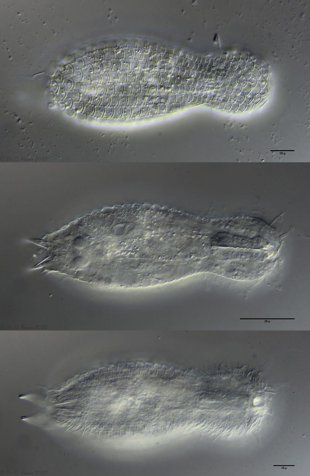
One of the animals found had reached its postpathenogenetic (hermaphrodite) phase and was already carrying an X organ and sperm bundle:
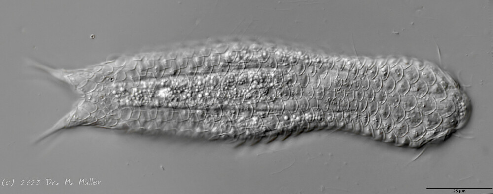
L. minor chaetifer: above dorsally with smooth scales typical of Lepidodermella;
middle in cross section with X-organ and sperm bundle;
below ventral scaling with distinct terminal plates ( Ole Riemann).
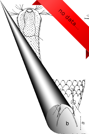
In Sima Marsh the somewhat divergent subspecies Lepidodermella minor chaetifer was found, differing from the species L. minor minor by a pair of spines at the base of the toes:
L. minor chaetifer: the pair of spines at the base of the toes is characteristic of the subspecies chaetifer
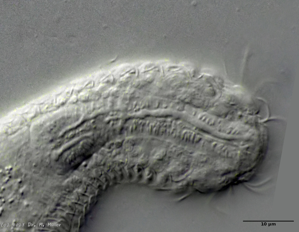
Another striking feature of this species is the single tooth projecting asymmetrically from the oral cavity:
Dorsal scales: 7-9 rows of 25-30 very differently shaped, spikeless, strongly overlapping scales; head and trunk scales elongated oval with triangular / broadened front end (12 x 10 µm); neck scales transversely oval, short (5 x 9 µm); hypostomium: two cusps
Ventral scales: ventral intermediate field with more than 20 transverse claspers below pharynx and 3-5 rows of quadrangular rounded, overlapping scales; 2 large terminal plates and some small, round scales; ciliary bands anteriorly mostly united
Oecology: Frequent, found everywhere
Particularities: well delimited by rectangular scale plates and large terminal plates; very variable and well studied species
Lepidodermella squamata is one of the best-studied gastrotrichs, as it is kept in culture in many laboratories around the world. However, the “cultivated forms” often differ significantly from the “wild forms”, as the animals, with their enormous generation rates of around 50 per year, can react quickly to the adaptive pressure of the habitat. L. squamata is therefore considered a very variable species. The photos shown here are of wild forms.
Dorsal scales
Ventral scales; large terminal plates; rectangular braces under neck
L. squamata is one of the many species of gastrotrichs in which the glandular ducts on the surface of the pharynx are clearly visible. These ducts connect individual gland cells in the pharyngeal wall and channel the secretions of the glands into a common excretory duct that pours into the animal’s oral cavity:

 L. minor chaetifer: above dorsally with smooth scales typical of Lepidodermella;
middle in cross section with X-organ and sperm bundle;
below ventral scaling with distinct terminal plates ( Ole Riemann).
L. minor chaetifer: above dorsally with smooth scales typical of Lepidodermella;
middle in cross section with X-organ and sperm bundle;
below ventral scaling with distinct terminal plates ( Ole Riemann).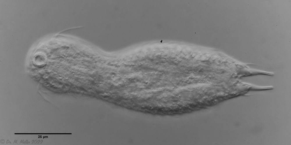 L. minor chaetifer: the pair of spines at the base of the toes is characteristic of the subspecies chaetifer
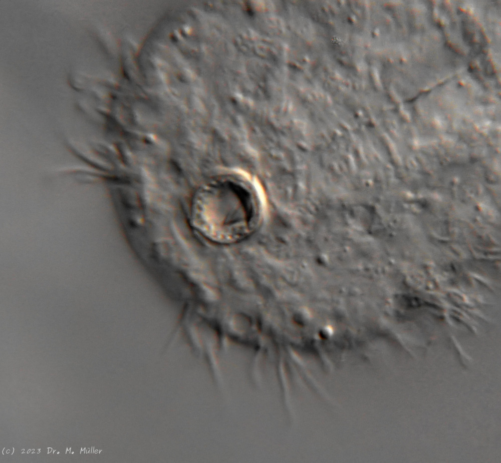
 Dorsal scales
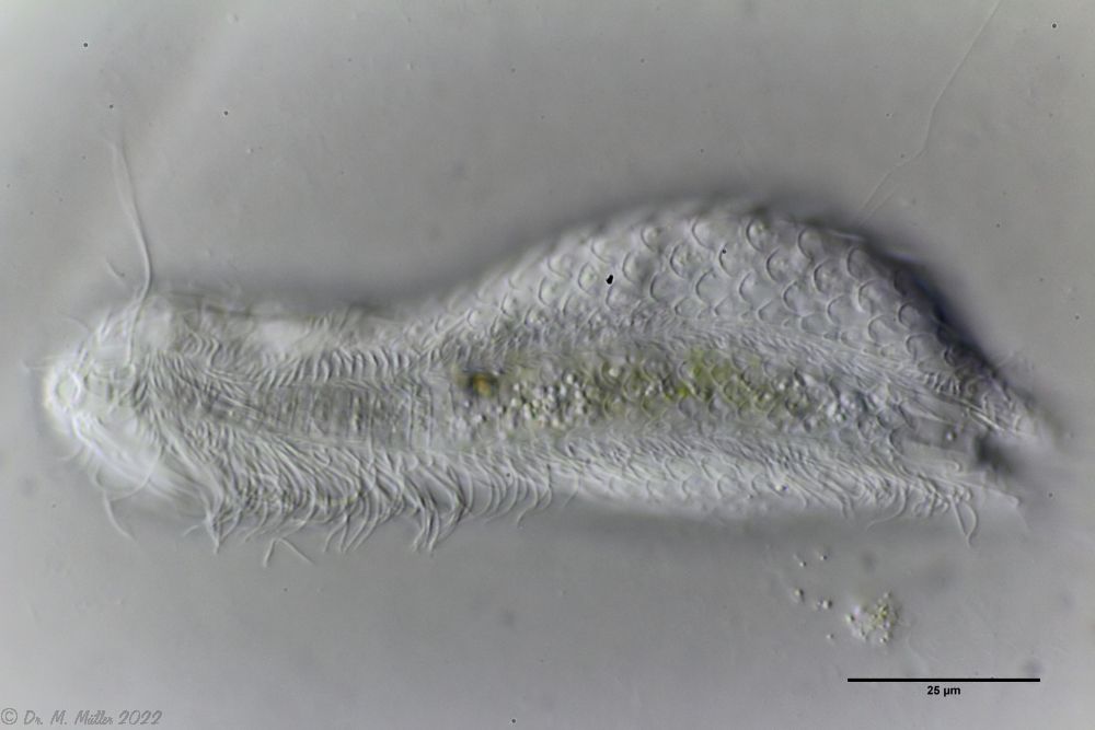 Ventral scales; large terminal plates; rectangular braces under neck