
Heterolepidoderma macrops
110 µm - 150 µm
Width:
30 µm
Width of the head ( five-lobed ):
25 µm - 28 µm
µm
Length of the furca:
15 µm - 16 µm
Adhessive tubes (thin, terminal thickened):
67% of furca
Pharyx ( cylindrical, terminal swollen ):
36 µm - 38 µm
Diameter of the mouth ( around ):
4.5 µm
Dorsal scales:
22-27 rows, each with 25-40 thin, elliptical keel scales (2 µm long) without terminal spine; toes unscaled
Ventral scales:
two small keeled terminal plates, otherwise naked
Oecology:
sehr widespread
Similar species:
H. ocellatum : larger, fewer scales; scaled toes and Ventral intercilliary field
Particularities:
mostly relatively large pseudocells
Fundorte:
Heterolepidoderma macrops is one of the most common and widespread Heterolepidoderma species and was certainly often confused in the past with H. ocellatum from which it differs - apart from some details that are difficult to see - mainly by the larger number of scales:
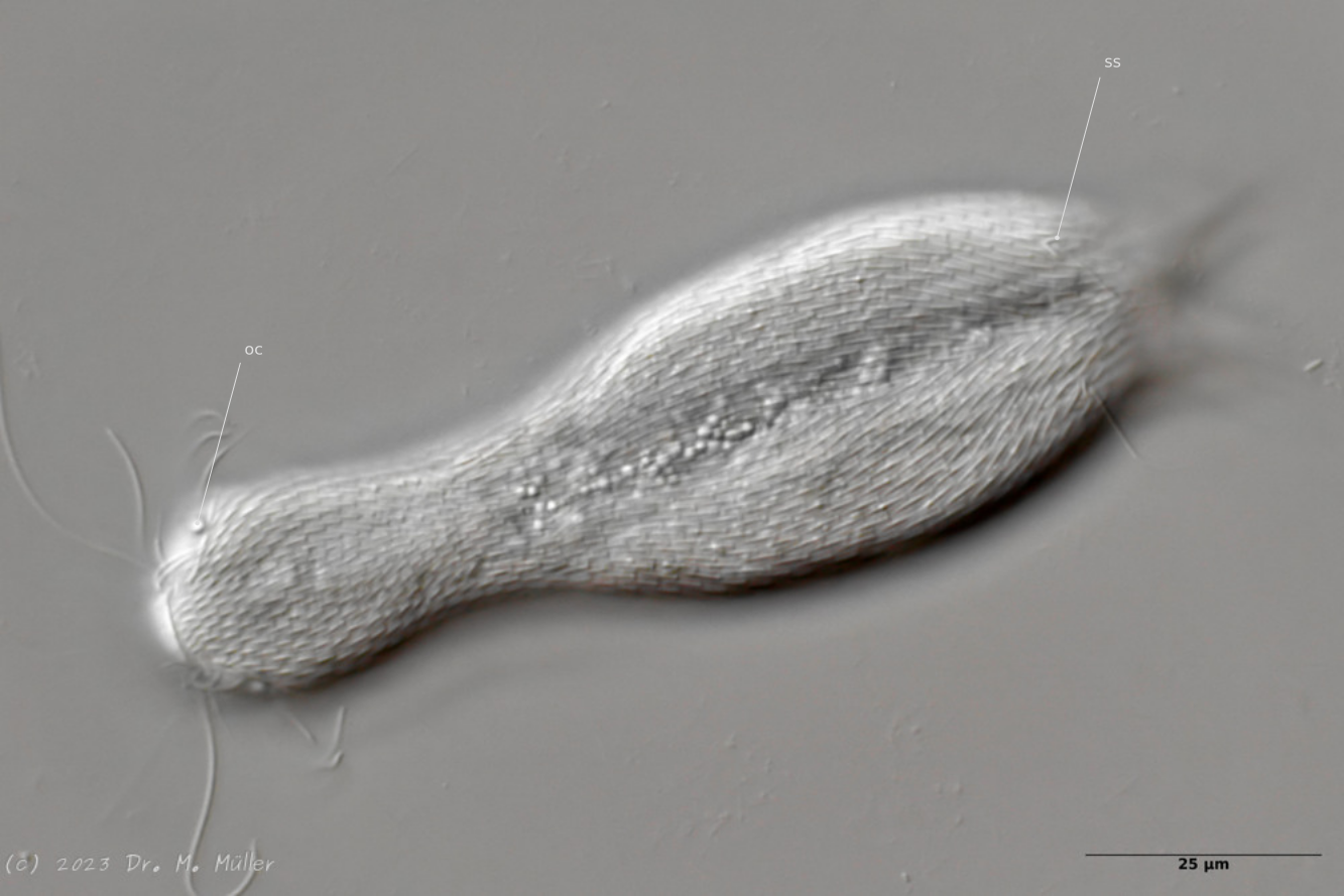
Dorsal view; oc: pseudocells; ss: sensory scales.
In most animals, the pseudocells resembling eyes are very conspicuous, but they have no sensory function. The posterior sensory hairs are located on small, triangular “sensory scales”.
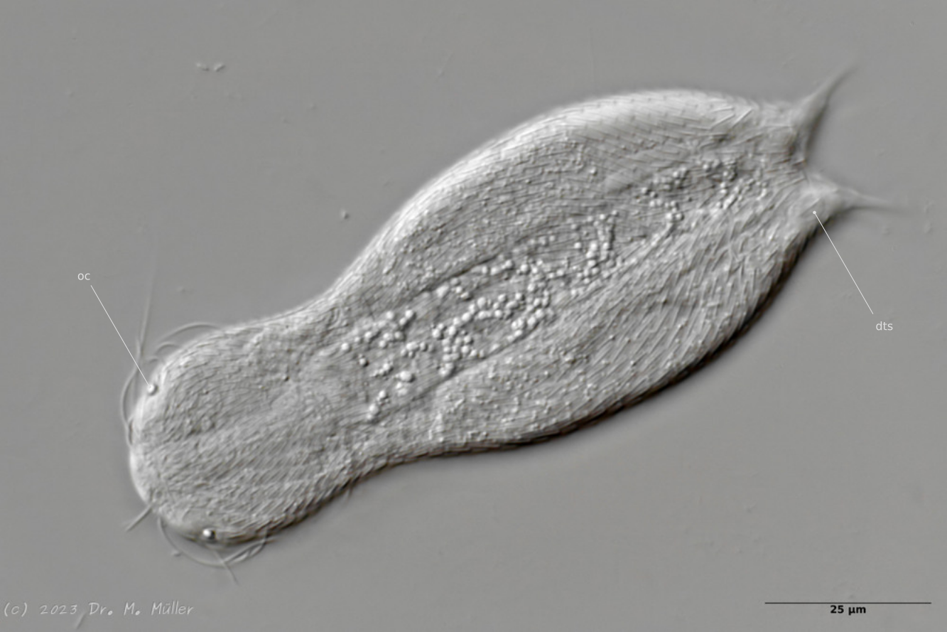
Dorsal with focus on the dorsal terminal scales (dts).
The toes of the animals are unscaled. The scale ends with a pair of relatively large keel scales at the base of the toes.
*Cross section
In the section you can see that the small scales - in contrast to H. ocellatum - do not bear a distal process. The pharynx is slightly swollen on both sides.
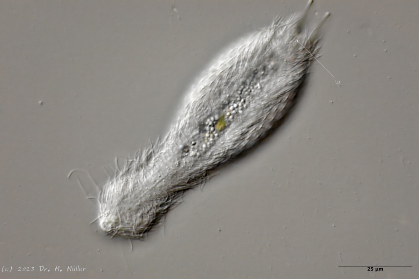
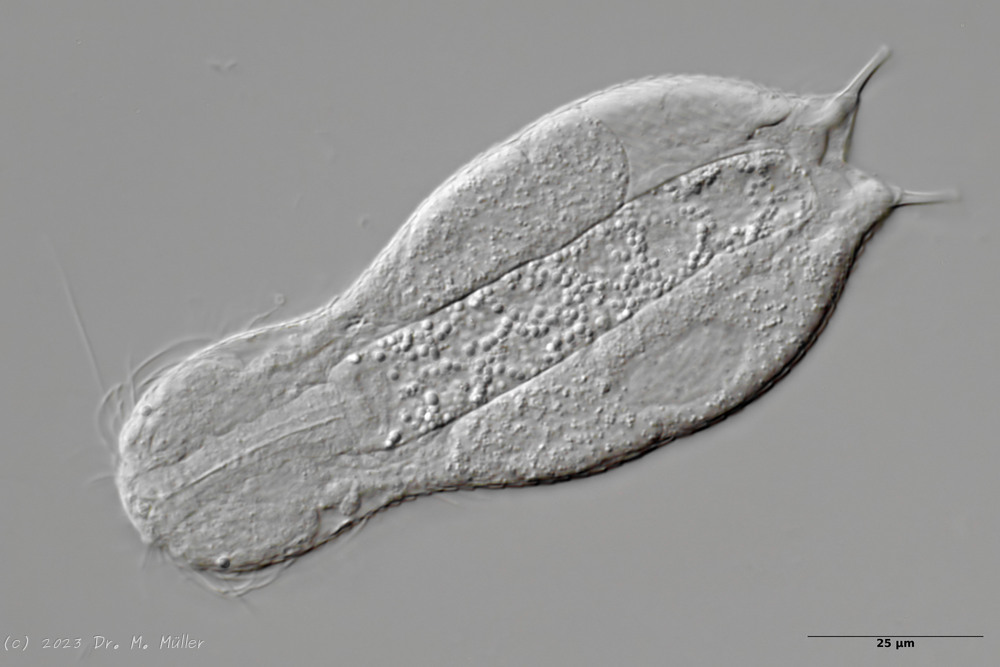
Central view with terminal plates (tp)
Ventrally, the animal bears only a pair of small, oval terminal plates. The remainder of the ventral interfield is unscaled. The adhesive tubes are thickened terminally.
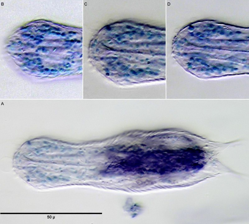
Staining of nuclei with methyl green; B C D: Nuclei of the brain; A: overview.
If H. macrops is stained with a selective nuclear dye like methyl green, only the nuclei of the animals are stained. This gives a good overview of the structure of the brain. Clearly visible in subfigure B is the nucleus-free area dorsal to the pharynx, which is referred to by (Zelinka, 1889) and (Remane, 1936) as the “dorsal commissure”, i.e. a nucleus-free connection of the two cerebral hemispheres made of nerve fibers, and which is also confirmed in more recent studies (Schmidt-Rhaesa and Holger Rothe, 2014) . Lowering the focus to the midplane of the pharynx (subimage C), one recognizes the pharynx consisting of a few cells surrounded on both sides by brain cells. Ventrally, no connection of the two cerebral hemispheres can be seen below the pharynx. Thus, the brain does not completely enclose the pharynx and delimits the animals to the Cycloneuralia. The already unstained conspicuous pseudocells prove to be stainable with methyl green - a strong indication that these structures are the prominent nuclei of the sensory neurons of the tactile hairs.
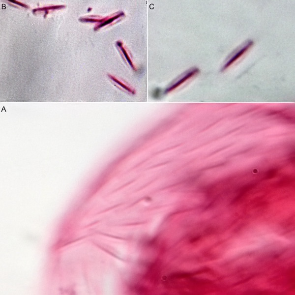
Scale preparation with eosin.
The scale shape of the small dorsal keel scales of H. macrops cannot be judged exactly in the scale compound on the living animal. A more precise [scale preparation](/technique/scale preparation) clearly shows the oval shape of the scales, which - in contrast to H. ocellatum - do not wear a distal tip.
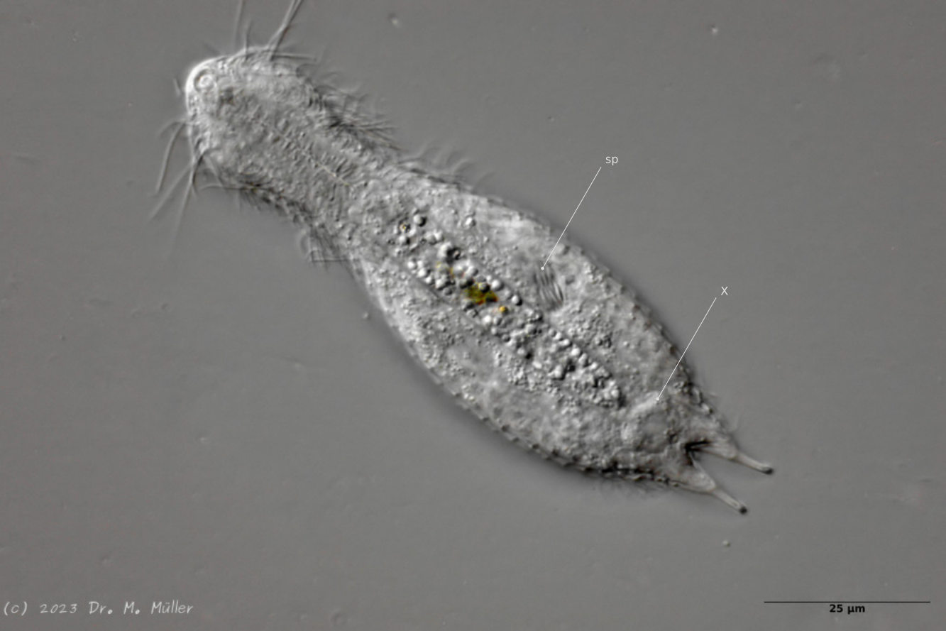
Postparthenogenetic (hermaphroditic) phase; sp: sperm bundle; X: X-organ.
After a phase with parthenogenetic reproduction, the animals enter the so-called “postpathenogenetic phase”. In this stage of life, the animals reform into hermaphrodites and simultaneously develop eggs and sperm produced in bundles ventrally to the side of the intestine - they reform into true hermaphrodites.
In H. macrops the spermatoids are spindle-shaped. At the same time, a bipartite, so-called X-organ with unknown function is formed. Here the X-organ consists of two spherical, secretion-filled cysts connected by a bar (or connecting duct).
(Kisielewski, 1981)